 The rats were fed an ordinary rat chow found to contain GMOs on PCR analysis using probes for the cauliflower mosaic virus 35S promoter, a gene control element in more than 80 % of commercial GM crops grown with potential health hazards predicted since 1999.
 Dr Mae-Wan Ho
Institute of Science in Society
Researchers led by Hanaa Oraby at Egypt’s National Research Center in Cairo are not the first to look for horizontal transfer of genetically modified (GM) DNA into animal cells, but certainly among the first to do an experiment aimed at detecting it and succeeded [1]. Horizontal gene transfer is the direct uptake of DNA (or RNA) into cells and integration of the sequence into the cell’s genome. Some of us regard horizontal gene transfer as the most serious hidden hazard of genetically modified organisms (GMOs) released into the environment ([2] Horizontal Gene Transfer – The Hidden Hazards of Genetic Engineering (ISIS special report). But a prevailing culture of denial by vested interests and regulators has obstructed proper investigation until very recently (see [3] Horizontal Transfer of GM DNA Widespread, SiS 64).
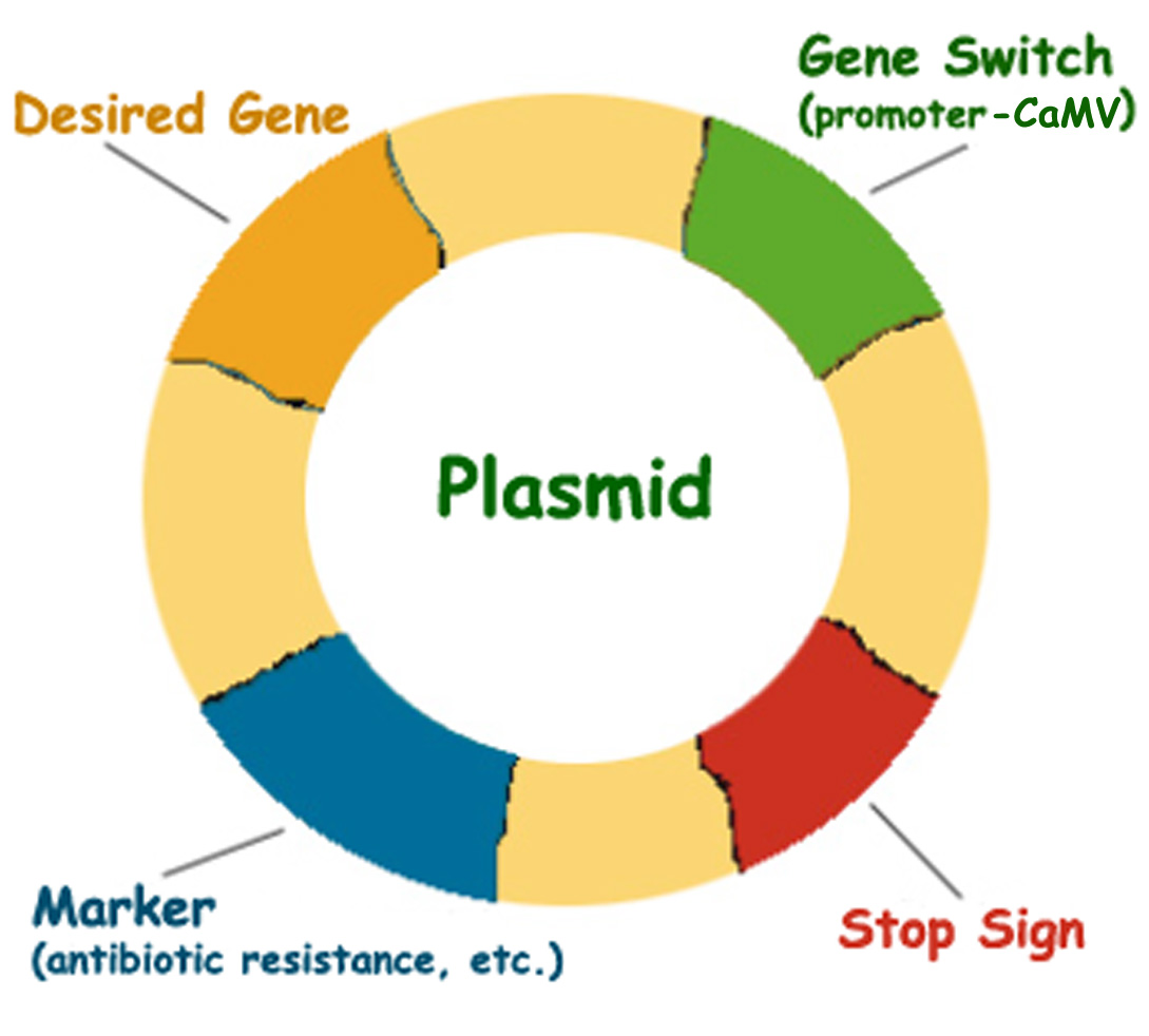
A GMO is an organism with synthetic foreign DNA gene sequences inserted into its genome in a laboratory process of artificial genetic modification that bypasses normal reproduction. Part of the foreign DNA is a control element called a promoter that is necessary for expressing the foreign genes. The most widely used is the cauliflower mosaic virus (CaMV) 35S promoter (which is what enables the virus to hijack the cell for making endless copies of the virus). The CaMV 35S promoter is now in more than 80 % of all GM plants [4], and is the first test for the presence of GMOs in unknown samples.
Probing for CaMV 35S promoter in the rat diet and in rat tissues
The Cairo researchers used three pairs of primers – specific short anchoring sequences that bind by specific base-pairing to the opposite ends of the DNA segment of interest – so as to amplify different segments from the CaMV 35S promoter with PCR (polymerase chain reaction). The amplified segments can then be isolated and detected on electrophoresis. The primers together amplify nearly 80 % of the entire promoter sequence. The experimental diet was an ordinary lab chow containing 60 % yellow maize and 34 % soybean, but unlabelled as to whether it is GM or not. The presence of GM material in the diet was ascertained using PCR assay with the three pairs of primers, which all gave the expected positive results, indicating that the diet contained GM material (up to a maximum of 94 %, if both the soybean and maize were completely GM).The experimental animals consisted of 29 male Wistar albino rats immediately after weaning (age three to four weeks), which were divided into two main groups. One was fed on the lab chow containing GM ingredients for three months, and were further divided into three subgroups of 5, 5, and 7 animals, euthanized after 30, 60, and 90 days. The other control group was fed on a balanced non-GM diet for the same period and euthanized at the end of the experiment.
The lab chow diet used all through this experiment gave positive results when screened with primers for CaMV 35S promoter. The expected 195 bp (base pairs) amplified product was detected in all samples of the GM diet, but was absent in the control diet made up with non-GM material to match the nutritional content of the GM diet. As further confirmation, the PCR product obtained from the GM diet was sequenced, and shown to have 100 % identity with the CaMV 35S promoter at nucleotide coordinate 7190-7380 of the CaMV on sequence alignment analysis using the GenBank database. It also showed 100 % sequence identity with a number of binary vectors used for transferring genes in the lab that also contain the CaMV 35S promoter.
PCR analysis of the different tissues showed amplified sequence segments of the expected sizes for the three pairs of primers – 70, 88 and 195 bp – in some of the DNA samples of blood, liver and brain of rats fed the GM diet after 30, 60 and 90 days. None of the three primers gave amplification product in DNA samples of tissues from the controls fed the non-GM diet.
The 195 bp segment amplified from DNA samples of liver and brain in rats fed GM diet was subjected to DNA sequencing, and comparison with GenBank database revealed 100 % identity with the CaMV whole genome at the same nucleotide coordinates 7190-7384 for the 35S promoter. Furthermore, it also showed 100 % identity with the PCR segment amplified from the GM diet, and with the binary vectors segments that are 100 % identical to the PCR product from the GM diet.
Feeding rats with GM diet for 30, 60, and 90 days increased the mean transfer frequency of GM target sequences significantly from 0 in the controls to 8 + 0.0000 %, 12.3 + 1.2018 % and 16.7 + 2.4529 % respectively. Thus, there is a cumulative effect with time of exposure.
Bearing in mind that the three primer pairs together amplify nearly 80 % of the entire CaMV 35S promoter, and in some animals, the three segments were all amplified in the same sample, it suggests that the whole CaMV 35S promoter may have been transferred into the genome of those animals. Considering that even a promoter containing only 46 bp of the 5’ sequence from the CaMV 35S promoter was previously reported to be sufficient for accurate initiation of gene transcription for gene expression [5], it is highly likely that the transferred CaMV35S promoter sequence would alter the activity of some genes in the host cell genome that may have harmful consequences (see later).
There was no significant difference in the rate of transfer into blood, liver, or brain tissues. Moreover, the frequency of uptake for the larger segments was greater than that for the smaller segments; thus, the transfer frequency of the 70 bp segment was the lowest at 1.09 + 0.4161 %, the 88 bp at 2.09 + 0.7318 %, and the 159 bp at 3.8 + 0.8069 %. This finding is consistent with previous researchers who postulated that the shorter the fragment, the lower the uptake efficiency [6].
GM fed rats suffered damages to liver, kidney and testis
In a separate report written by some of the same researchers in Cairo, the same GM diet was fed to identical rats in a post-market safety assessment of GMOs [7]. Biochemical, histopathological, and cytogenetic analyses on liver, kidney, and testis revealed that the GM diet fed for 30, 60 and 90 days suffered significant deleterious effects.A total of 30 rats were fed the GM diet, 10 each for 30 days, 60 days and 90 days. The controls were on a wheat-based non-GM nutritionally matched diet for similar periods of time.
Alanine aminotransferase (ALT) and aspartate aminotransferase (AST) enzyme activities were measured in blood serum as indicators of liver cell damage. Creatinine and uric acid levels were determined also in blood serum as indicators of kidney function. Malondialdehyde in liver cells resulting from lipid peroxidation is a biomarker of oxidative stress. In addition, specimens of liver, kidney and testis were dissected immediately after the rats were euthanized and sectioned for histopathological and histochemical investigations. Cytogenetic and DNA damage analyses were carried out on testis and liver. Chromosome analysis was done on germ cells from the testis. Sperms were examined for morphological abnormalities and DNA fragmentation was determined in liver cells.
The results were unambiguous.
Histopathology of liver, kidney and testis
Liver cells showed slight damage in rats fed GM diet for 30 days, with damage increasing after 60 and 90 days. The effects start as a slight dilatation and congestion of the central vein (supplying the liver) and fragmentation of the nucleus in some cells. After 60 days, mild cellular infiltration (from the blood) was observed. After 90 days, the blood sinusoids (spaces) also showed slight dilatation and congestion (Figure 1).
Figure 1 Liver sections: a, control; b, 30 day GM-fed; c, 60 day GM-fed; d 90 day GM fed (see text for details)
Kidney sections show damaging effects of the GM diet evident even after the first 3o days as interstitial haemorrhage (blood in spaces between cells) and a widening of the tubules. This got worse in 60 and 90 days (Figure 2).
Figure 2 Kidney sections: a, control; b, 30 day GM-fed; c, 60 day GM-fed; d 90 day GM fed (see text for details)
The testis of rats fed on GM food for 30 days showed mild thickening of the basement membrane of the seminiferous tubules (where germ cells develop) with gaps appearing between the germinal epithelium of some tubules. At 60 and 90 days, an increase in the connective tissue component and in the number of Leidig cells (in the connective tissue), with a disarrangement of the germinal epithelium (Figure 3).
Figure 3 Testis sections: a, control; b, 30 day GM-fed; c, 60 day GM-fed; d 90 day GM fed (see text for details)
Protein content in liver tissues decreased significantly after 30 or 90 days, indicating dysfunction of some hepatocytes. Abnormal cellular activity in the kidney was also confirmed by a statistically significant increase in the protein content.Consistent with the liver and kidney damages seen, AST and ALT activity increased in the serum of experimental rats by 33 to 107 % and 33 to 92 % respectively. Blood creatine and uric acid concentrations significantly increased by 15 to 315 % and 37 to 96 % respectively. MDA concentrations in liver, as an indicator of oxidative stress, increased significantly in all animals fed GM diet by 286 to as high as 940%.
Mitotic index (as a measure of cell division) was significantly reduced in the 60 and 90 days fed rats from 8.8 + 0.326 % in controls to 8.4 + 0.221 % (30 days), 6.8 + 0.466 % (60 days) and 6.6 + 0.266 % (90 days). Concomitantly, there was a significant increase in chromosomal aberrations, from 0.4 + 0.163 % in controls to 6.6 + 0.221 %, 13.8 + 0.326 % and 8.0 + 0.632 % respectively for 30, 60 and 90 day GM fed rats. The frequency of morphologically abnormal sperm increased by up to two-fold, from 3.33 +0.35 % to 5.83 + 0.60 %, 7.8 + 0.65 % and 6.6 + 0.24 %. At the same time DNA fragmentation went up from 11.83 + 0.7 % in controls to 19.0 + 1.2 %, 28.3 + 1.6 % and 24.3+ 0.7% respectively.
The researchers concluded the results [7] “indicate that there are health hazards linked to the ingestion of diets containing genetically modified components.”
However, the investigations have not gone further into the mechanisms whereby the genetically modified components were hazardous to health; nor do the results directly implicate the CaMV 35S promoter found transferred to rat blood, liver and brain in the other report [1]. It will be useful to review the potential hazards of the CaMV 35S promoter, which were first pointed out 15 years ago.
Predicted hazards of cauliflower mosaic virus 35S promoter
When first deployed, geneticists assumed that the CaMV 35S promoter would only work in plants, as the complete virus (wrapped in its protein coat) specifically infects only plant cells. But it soon transpired that the isolated piece of promoter DNA without its coat is extremely promiscuous, and works in cells across kingdoms of plants and animals, as well as bacteria. We issued a serious warning against its use in 1999 [8] Cauliflower Mosaic Viral Promoter – A Recipe for Disaster (ISIS scientific publication) when it was found to have a recombination hotspot where it tends to fragment and join, which makes it prone to unintended (horizontal) gene transfer into cells of all organisms exposed to the GMO, including bacteria, fungi, pollinators, wild animals and humans (see [9] CaMV 35S promoter fragmentation hotspot confirmed, and it is active in animals (ISIS scientific publication). What that implies is the CaMV 35S promoter can break loose from the plant genome DNA and jump into the genome of all those other cells, with the potential to mutate, activate or inactivate genes (including those leading to cancer), reactivate dormant viruses, or create new viruses by recombination (gene shuffling) [8, 10] (Hazards of Transgenic Plants Containing the Cauliflower Mosaic Viral Promoter, ISIS scientific publication) But our warnings were met with abuse and denial and ultimately ignored.Since then, evidence has emerged that the CaMV 35S promoter may enhance the multiplication of disease-associated viruses including HIV and cytomegalovirus through the induction of proteins required for transcription of the viruses [11] (New Evidence Links CaMV 35S Promoter to HIV Transcription, ISIS scientific publication). Further, the CaMV 35S promoter overlaps with a virus gene, the product of which is toxic to plants and likely also to animals [12]. For a more detailed description on the risks of the CaMV 35S promoter and indeed on GMOs in general, see [13] and final chapter in [14] Ban GMOs Now, ISIS Report.
To conclude
GMOs are once again found to be deleterious for health in a feeding trial that last no longer than 90 days. And within that time, the most widespread piece of transgenic DNA found in the GM diet, the CaMV 35S promoter, was found transferred horizontally into the animals’ tissues at high frequencies. The CaMV 35S promoter is not the only hazardous piece of transgenic DNA, there are similar aggressive promoters designed to make genes express out of context, as well as genes coding for antibiotics and other dangerous functions, together with numerous recombination hotspots that enhance horizontal gene transfer; all of which contribute to making all GMOs unsafe. That is indeed the conclusion from research carried out by scientists independent of the industry up to now, which fully corroborates what farmers have been witnessing in their livestock and doctors in their patients for years [14]. People need to take immediate action to ban GMOs from their own home and local communities. Governments should recall all GMOs from the market. And companies and regulators should face prosecution for causing damages to health and criminal negligence.A fully referenced and illustrated version of this article is posted on ISIS members website and otherwise available for download here
If you find this report useful, please support ISIS by subscribing to our magazine Science in Society, and encourage your friends to do so. Or have a look at the ISIS bookstore for other publications
Source: http://farmwars.info/?p=13753